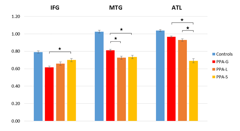
Figure 1. Normalized cortical volume in each ROI.
All subtypes showed reduced volume, in all ROIs, compared to the control group. Asterisks indicate significant differences in volume between PPA groups (Bonferroni corrected). IFG = Inferior Frontal Gyrus; MTG = Middle Temporal Gyrus; ATL = Anterior Temporal Lobe. PPA = Primary Progressive Aphasia; PPA-G = Nonfluent/agrammatic variant of PPA; PPA-L = Logopenic variant of PPA; PPA-S = Semantic variant of PPA.

