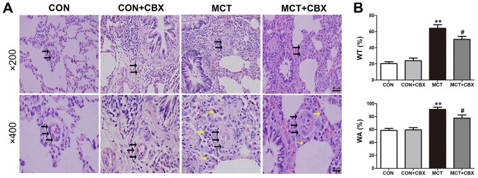
Figure 2.
MCT-induced remodeling of small pulmonary arterioles and inflammatory cell infiltration is alleviated by CBX. (A) Representative hematoxylin and eosin staining demonstrating histopathological changes in small pulmonary arterioles at magnification, ×200 (scale bar=50 µm; upper panel) and magnification, ×400 (scale bar=25 µm; lower panel). Pulmonary arteriole walls in the control group were thin and single layered. The intima, media and externa were difficult to distinguish. Pulmonary vascular remodeling was observed in MCT-treated animals, characterized by obliterated vessels (black arrows); no obliteration was detected in the control rats and the rats treated with a single intraperitoneal injection of CBX. CBX prevented pulmonary arterial obliteration in small pulmonary arterioles of MCT-treated animals. The black and yellow arrows indicate pulmonary arterial obliteration and perivascular infiltration of inflammatory cells in small pulmonary arterioles, respectively. (B) WT% and WA% of the MCT-treated rats were significantly increased, and CBX treatment of the MCT rats significantly decreased WT% and WA%. Data are presented as the mean ± standard error of the mean of 6 rats/group. **P<0.01 vs. control. #P<0.05 vs. MCT group. CBX, carbenoxolone; MCT, monocrotaline; CON, control; WT%, percentage of vascular wall thickness; WA%, percentage of the vascular wall area.

