Figure 4.
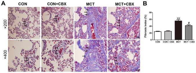
Administration of CBX ameliorates MCT-induced pulmonary fibrosis. (A) Masson trichrome staining of lung tissue at magnification, ×200 (scale bar =50 µm; upper panel) and magnification, ×400 (scale bar =25 µm; lower panel). Black arrows indicate collagen deposition, indicative of fibrosis, and collagen fibers are stained blue. Nuclei are stained dark red/purple, and the cytoplasm is stained red/pink. (B) Quantification of interstitial fibrosis as a percentage derived from positive collagen staining. The lung fibrosis index was defined as the percentage of the total area stained with collagen divided by the sum of total connective tissue area in the entire visual field. Data are presented as the mean ± standard error of the mean of 6 rats/group. **P<0.01 vs. control. #P<0.05 vs. MCT group. CBX, carbenoxolone; MCT, monocrotaline; CON, control.

