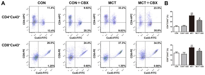
Figure 9.
CBX administration decreases the percentages of Cx43-expressing CD4+ and CD8+ T cells in the lung tissues of the MCT-treated rats. (A) Representative flow cytometry plots and (B) quantitative analysis demonstrating the proportions of CD4+Cx43+ or CD8+Cx43+ double-positive T cells were increased in lung tissues of MCT-treated rats compared with the control group. There was a significant decrease in the frequencies of CD4+Cx43+ and CD8+Cx43+ T cells in the MCT + CBX-treated rats compared with the MCT-treated rats. The frequencies of CD4+Cx43+ and CD8+Cx43+ T cells did not significantly differ between the lungs of control rats and the rats treated with a single intraperitoneal injection of CBX. Data are presented as the mean ± standard error of the mean of 6 rats/group. **P<0.01 vs. the control group. #P<0.05 vs. the MCT-treated rats. APC, allophycocyanin; CBX, carbenoxolone; CON, control; Cx, connexin; CD, cluster of differentiation; FITC, fluorescein isothiocyanate; MCT, monocrotaline.

