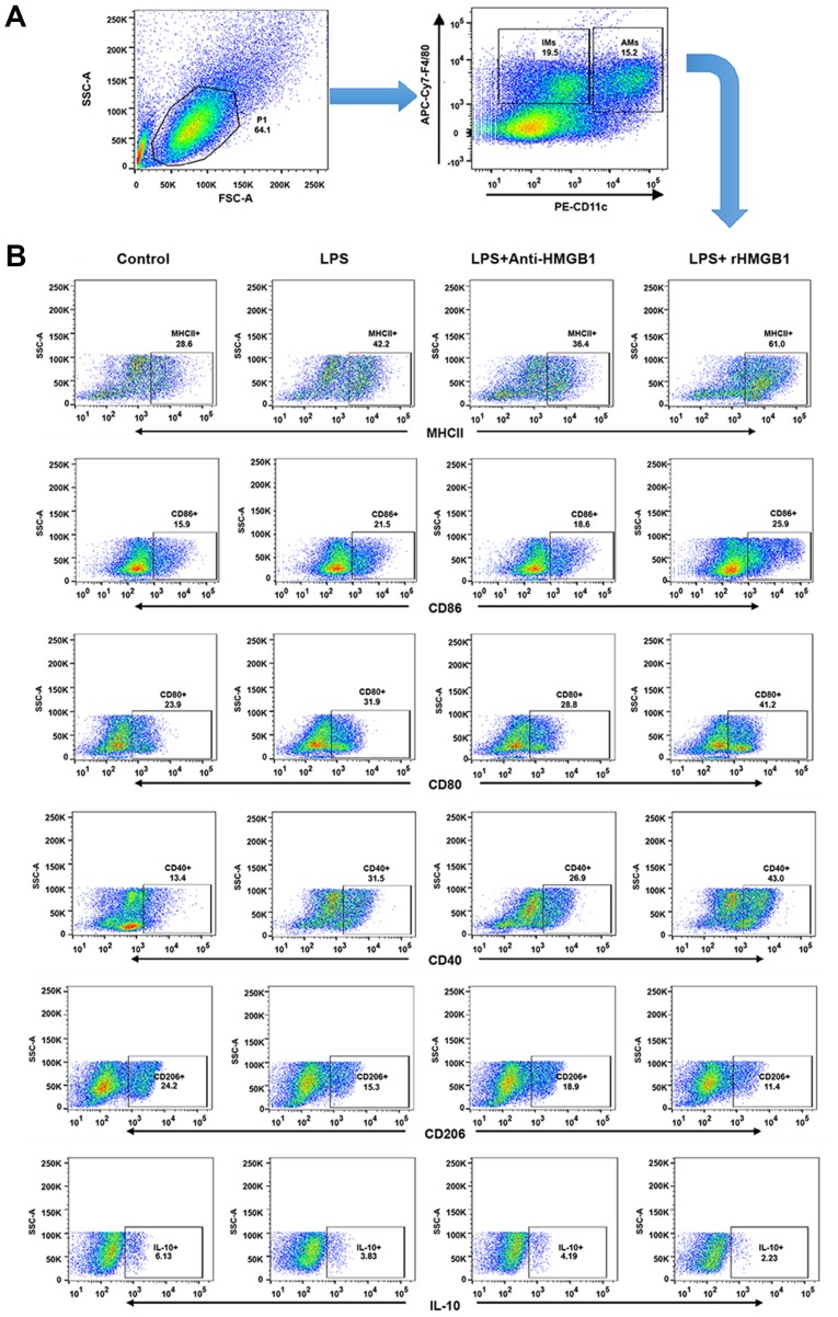
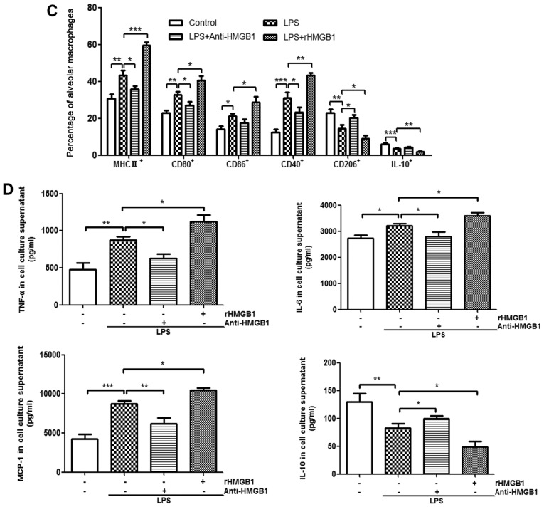
Figure 3.
Upregulation surface markers and cytokines of M1 macrophages by extracellular HMGB1 (A) Flow cytometry was used to identify F4/80+ and CD11c+ lung AMs isolated from lung MNCs of mice. IMs were marked as F4/80+ and CD11c− MNCs. (B) Detection of expression levels of MHC II+, CD80+, CD86+, CD40+, CD206+ and IL-10 on AM cell surface. All experiments were repeated more than three times (n=4-6 mice per each group). Data presented is from a representative experiment. All data are expressed as the mean ± standard deviation. Upregulation surface markers and cytokines of M1 macrophages by extracellular HMGB1. (C) The percentages of lung AMs expressing MHC II, CD80, CD86, CD40, CD206 and IL-10 were calculated. (D) AM activation is defined by two distinct polarization states: M1 and M2. M1-related cytokines (tumor necrosis factor-α, IL-6, and MCP-1) and M2-related cytokine (IL-10) were detected in culture supernatant of BMMs by ELISA. All experiments were repeated more than three times (n=4-6 mice per each group). Data presented is from a representative experiment. All data are expressed as the mean ± standard deviation. *P<0.05, **P<0.01 and ***P<0.001 vs. LPS group. LPS, lipopolysaccharide; IL, interleukin; MCP, myeloperoxidase; rHMGB1, recombinant High mobility group box 1; CD, cluster of differentiation; MHC, major histocompatibility complex; AMs, alveolar macrophages; MNCs, mono-nuclear cells; BMMs, bone-marrow derived macrophages.


