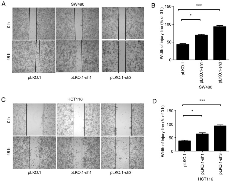
Figure 4.
Effects of siMyD88 on the migration of pLKO.1, pLKO.1-sh1 and pLKO.1-sh3 in the SW480 and HCT116 cells. (A) In the SW480 cells, representative images of pLKO.1, pLKO.1-sh1 and pLKO.1-sh3 cell wound healing and microscopic observations were photographed 0 and 48 h after scratching the cell surface. (B) The width of the scratch was analyzed using a histogram in the SW480 cells. (C) In the HCT116 cells, representative images of pLKO.1, pLKO.1-sh1 and pLKO.1-sh3 cells wound healing and microscopic observations were photographed 0 and 48 h after scratching the cell surface. (D) The width of the scratch was analyzed using a histogram in the HCT116 cells. Error bars represent mean ± SEM, representative of three experiments. *P<0.05, ***P<0.001.

