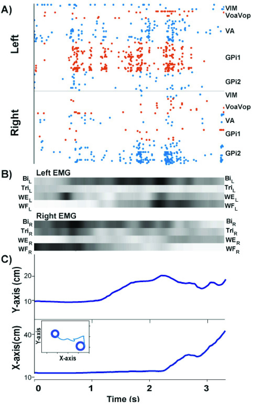
FIGURE 11.
Intracortical neural data synchronized with behavioral data collection in the EMU participant. The neural signals and synchronized kinematics are from a single discrete movement in the outward direction. A: Spike raster recorded from DBS electrodes in the left and right basal ganglia and thalamus. B: EMG signals from the left and right upper extremity displayed in scaled colormap format: the darker the color, the higher the signal intensity. C: Kinematics in the x- and y-direction on the MAGIC Table. The two-dimensional trajectory of the trial is displayed in the inset. Legend: VIM: ventralis intermedius medium. VoaVop: ventralis oralis anterior. ventral oralis posterior. VA: ventralis anterior. GPi: globus palidus internus. Bi: biceps, Tri: triceps, WE: wrist extension. WF: wrist flexor.

