Figure 1.

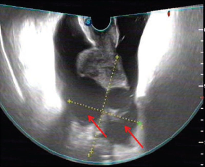
Gynecologic sonography showing an 8.8 × 7.5 cm mass (red arrow) with mixed density and irregular shape behind the uterus.

Gynecologic sonography showing an 8.8 × 7.5 cm mass (red arrow) with mixed density and irregular shape behind the uterus.