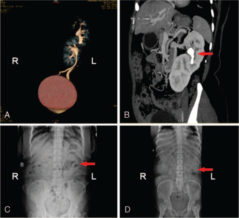
Figure 1.

3D computed tomography and X-ray images of patient 1. (A) 3D computed tomography revealed S-shaped right-to-left crossed-fused renal ectopia. (B) CT demonstrated the vascular anomaly and calculi in the left renal pelvis (arrow). (C) Preoperative abdominal X-ray revealed stone shadows (arrow) in the left abdominal area. (D) Postoperative KUB showed no stone shadows.
