Figure 1.

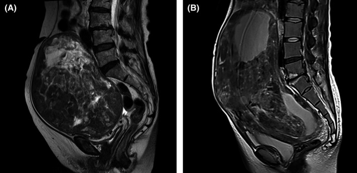
MRI for representative leiomyoma and leiomyosarcoma. Preoperative T2‐weighted MRI for a patient with uterine leiomyosarcoma (A) and leiomyoma (B). Both images were obtained from patients referred to National Cancer Center Hospital

MRI for representative leiomyoma and leiomyosarcoma. Preoperative T2‐weighted MRI for a patient with uterine leiomyosarcoma (A) and leiomyoma (B). Both images were obtained from patients referred to National Cancer Center Hospital