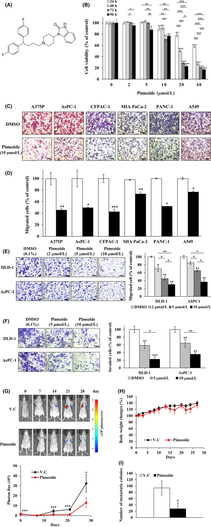
Figure 3.

Inhibition effect of pimozide on growth, migration, invasion, and metastasis. A, Chemical structure of pimozide. B, DLD‐1 cells were treated with pimozide (0, 2, 5, 10, 20 or 40 μM) for 24, 48, 72 or 96 h. Cell proliferation was measured by a WST‐1 assay (n = 3). C‐E, Cell migration assays were carried out with a Transwell system in different types of cancer cell lines. Migrated cells were stained with crystal violet and counted using the Image‐ProPlus 5.0 program (n = 3). Scale bars, 200 μm. F, Cell invasion assays were done using Matrigel‐coated Transwell inserts in DLD‐1 and AsPC‐1 cell lines and the migrating cells were quantified (n = 3). Scale bars, 200 μm. G, Inhibition of pancreatic cancer cell metastasis to the lungs by pimozide in the lung metastatic mouse model. Top, Representative images from luciferase‐expressing AsPC‐1 cells in the whole body (n = 6 per group). Bottom, Quantification of photon flux in the lungs at the indicated time points. V.C., vehicle control. H, Body weight was measured on each indicated day. I, On the 28th day, mice were killed and their lungs were dissected. Number of metastatic colonies in the lung was counted and averaged. (n = 6 per group). Data represent mean ± SD compared with the corresponding control, *P < .05, **P < .01, ***P < .001
