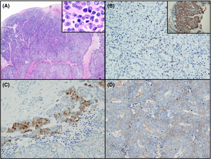
Figure 1.

Histological and immunohistochemical features of neuroendocrine carcinoma (NEC) in ascending colon. A, H&E staining shows that solid NEC have a high nuclear‐cytoplasmic ratio. B, Immunohistochemical staining shows that the nuclei of NEC stained positively for Ki‐67 in biopsy specimens (inset) and resected colon tissues. NEC stained positive for chromogranin A (C) and synaptophysin (D). Magnification: A, ×40; A inset, ×400; B, ×200; B inset, ×100; C and D, ×200
