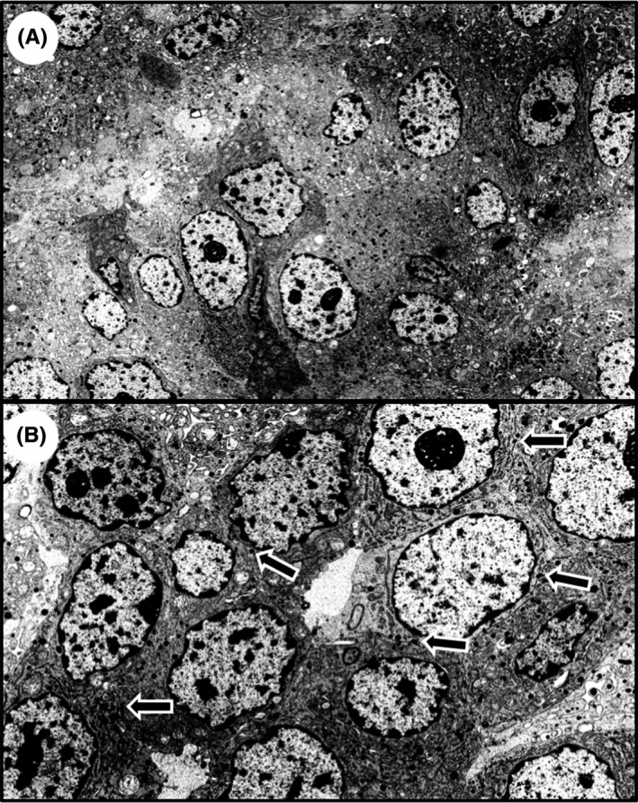
Figure 2.

Transmission electron microscopic (TEM) analysis of neuroendocrine carcinoma in ascending colon. TEM analysis shows neuroendocrine granules in the cytoplasm of the tumor cells (A), and desmosomes were observed at the tumor cell membrane (B, arrows). Magnification: A, ×4000; B, ×7000
