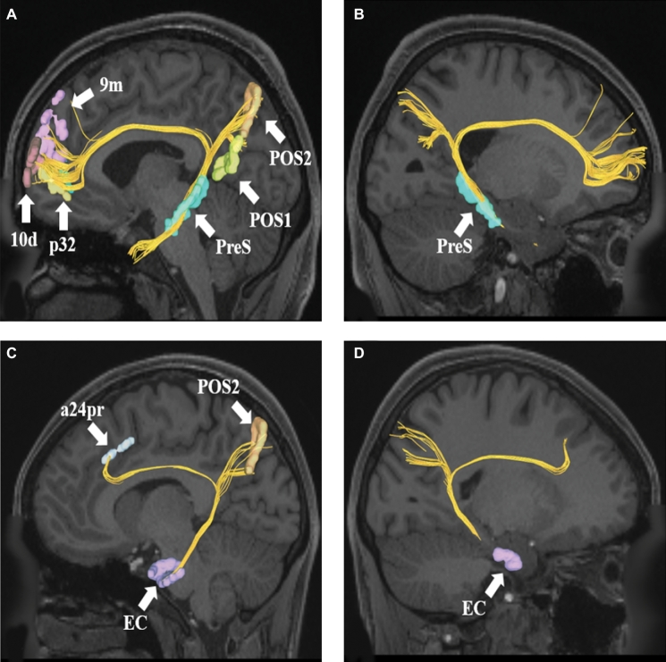
FIGURE 2.
A and B, Cingulate connections from area PreS in the left cerebral hemisphere. Connections are shown on T1-weighted MR images in the sagittal plane. Area PreS has connections to POS1, POS2, 9m, p32, and 10d in this subject brain. PanelBis the same image rotated 180° to demonstrate the precise location of PreS in the medial temporal lobe.CandD, Cingulate connections from area EC in the left cerebral hemisphere. Connections are shown on T1-weighted MR images in the sagittal plane. Area EC has connections to POS2 and a24pr in this subject brain. PanelDis the same image rotated 180° to demonstrate the precise location of EC in the medial temporal lobe.

