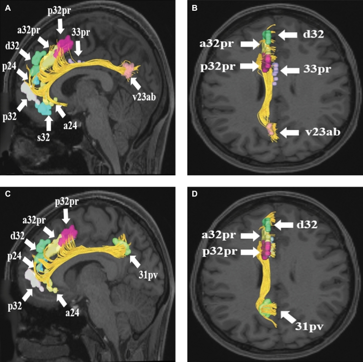
FIGURE 4.
A and B, Cingulate connections from area v23ab in the left cerebral hemisphere. Connections are shown on T1-weighted MR images in theA, sagittal andB, axial planes. Area v23ab has connections to a24, s32, p32, p24, d32, a32pr, p32pr, and 33pr in this subject brain.CandD, Cingulate connections from area 31pv in the left cerebral hemisphere. Connections are shown on T1-weighted MR images in theC, sagittal andD, axial planes. Area 31pv has connections to a24, p32, p24, d32, a32pr, and p32pr in this subject brain. All parcellations are identified with white arrows and corresponding labels.

