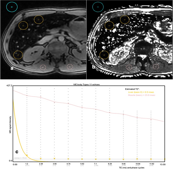
Fig. 5.
High LIC with 3-T imaging. a 2D ME-GRE sequence (first TE = 1.2 ms) obtained with body coil showing signal collapsed with a LIC of 521 μmol Fe/g as estimated by SIR method. In the same patient, pixelwise R2* map built by the 3D ME-GRE vendor solution (b), performed with surface coil, provides a wrong mean R2* of 188 s−1 which corresponds to slight iron overload (LIC = 59 μmol/g). The same patient was also scanned with another 3-T system from a different vendor (picture not provided) giving even a lower wrong R2* estimation by the 3D ME-GRE pixelwise map. R2* calculated using the same ROIs by MRQuantif (c) providing selected truncation fitting, excluding most of the points, was 1587s−1 corresponding to a LIC of 496 μmol Fe/g

