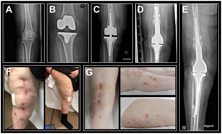
Figure 1.
Sequential anteroposterior radiographs of the patient's right knee indicate progression of bone loss and photographs of the right leg show persisting eczema. (A) Radiography prior to primary knee replacement depicts joint degeneration secondary to rheumatoid arthritis, 2009/03. (B) Pre-revisional radiograph indicates peri-implant bone loss and loosening of the tibial component, 2012/03. (C) Post-revisional radiograph depicts intense intraoperative usage of cement and progressive bone loss, 2012/07. (D) Radiographic status at the time of hypersensitivity diagnostics, 03/2018. (E) Latest radiographic status (patient in supine position), 01/2019. (F) Eczema at the time of hypersensitivity diagnostics, 03/2018. (G) Follow-up examination in the aftermath of rituximab therapy indicated an unimproved cutaneous status, 03/2019.

