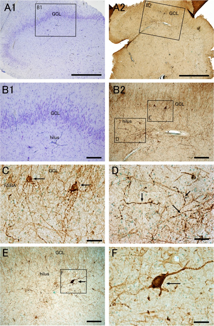
Figure 3.
Epileptic patients with granule cell dispersion and loss of hilar neurons have aberrant PSA-NCAM+ cells with multibasal dendrites in the hSGA, and unusually large PSA-NCAM+ cells and thick fibers with varicosities in the hilus. Nissl staining (A1,B1) and PSA-NCAM immunohistochemistry with methyl green nuclear staining (A2,B2,C–F) in the dentate gyrus of an epileptic patient (EP9, see Supplemental Table 1) with granule cell dispersion and loss of hilar neurons. The boxed regions in (A1,A2,B2,E) are enlarged in (B1,B2,C,D,F), respectively. Note the PSA-NCAM+ cells with multi-basal dendrites in the human subgranular area (hSGA) (B2,C), thick fibers with varicosities (B2,D), and strongly PSA-NCAM+ large cells (E,F) in the hilus. GCL, granule cell layer. Scale bars = 1 mm in A1 and A2; 200 μm in (B1,B2,E); and 50 μm in (C,D,F).

