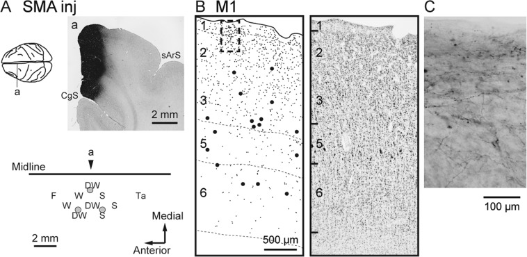
Figure 1.
Anterograde and retrograde labeling in M1 after BDA injections into SMA (monkey B). (A) Upper, Representative coronal section (a) through the injection site (right). CgS, cingulate sulcus; sArS, superior limb of the arcuate sulcus. The approximate anteroposterior level of the section (a) is indicated in the dorsal view of the brain (left). Lower, Result of intracortical microstimulation (ICMS) mapping. Filled gray circles denote the locations of the injection sites. In the somatotopic map, the body parts of which movements were evoked by ICMS are indicated as follows: D, digits; F, face; S, shoulder; Ta, tail; W, wrist. The combined letter, DW, represents the locus where different body-part movements were elicited in that order during microelectrode penetration. The arrowhead indicates the approximate anteroposterior level of the section (a) in the somatotopic map. (B) Representative coronal section showing the laminar organization of anterograde and retrograde labeling in M1 (left). Each small or large dot corresponds to one terminal varicosity or cell body labeled with BDA, respectively. Cortical layers are depicted on the left side of the section and demarcated with broken lines. A corresponding Nissl-stained section is shown on the right. (C) Higher-magnification photomicrograph showing BDA-labeled fibers, taken from the rectangular area in B.

