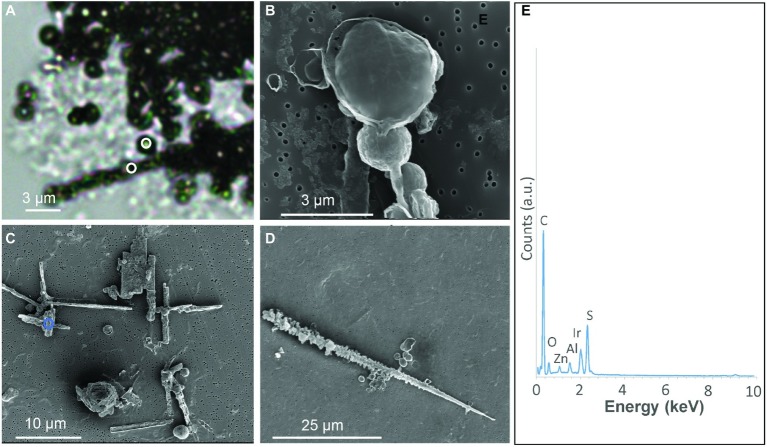
Figure 1.
S(0) particles formed in S. kujiense cultures with sulfide and thiosulfate. (A) Light micrograph of sulfur spheres and rods observed after 2 weeks of growth. White circles in (A) indicate where Raman spectra shown in Figure 2 were collected. (B) SEM image of spheres observed at 4 days. (C–D) SEM images of spheres and rods observed at 2 weeks. (E) XEDS spectrum collected on the area depicted by a blue circle in (C). Iridium (Ir) originates from the conductive coating of the SEM sample, while zinc (Zn) and aluminium (Al) are thought to originate from the sample holder and instrument.

