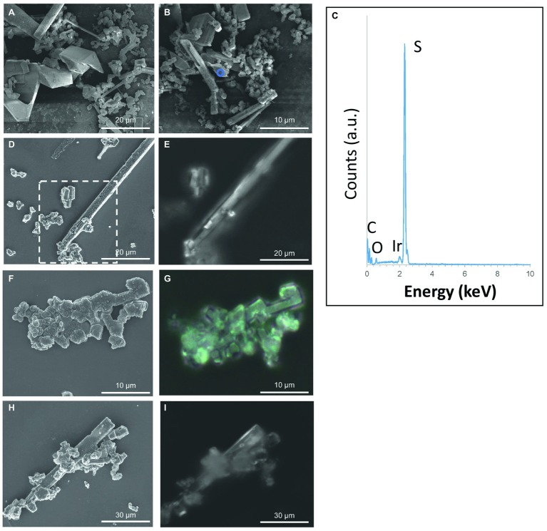
Figure 4.
S(0) particles formed in the (stationary phase) S. kujiense spent medium experiment. (A,B,D,F,H) SEM images. (C) XEDS spectrum collected on the area depicted by the blue circle in (B). (E,G,H) Correlative light microscopy images acquired with a Raman spectromicroscope on the areas corresponding to the white rectangle in image (D), and to images (F) and (H), respectively. Raman spectra were collected on the particles shown in (D–I) (see Figure 2). Note that an average of three Raman spectra were acquired on each particle, and that they were all similar to the representative spectrum shown in Figure 2.

