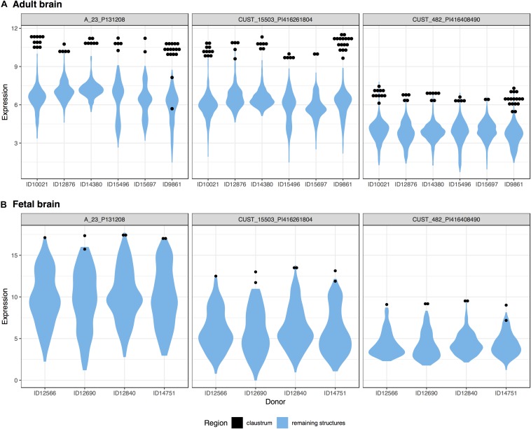
FIGURE 4.
Plots of NR4A2 expression in the adult (A) and fetal (B) brains. Expression (log2 intensity) is plotted on the y-axis for each of the three probes for NR4A2. Donor identification numbers are marked on the x-axis. Expression in the claustrum is marked in black, expression across the remaining brain regions is shown in blue violin plots.

