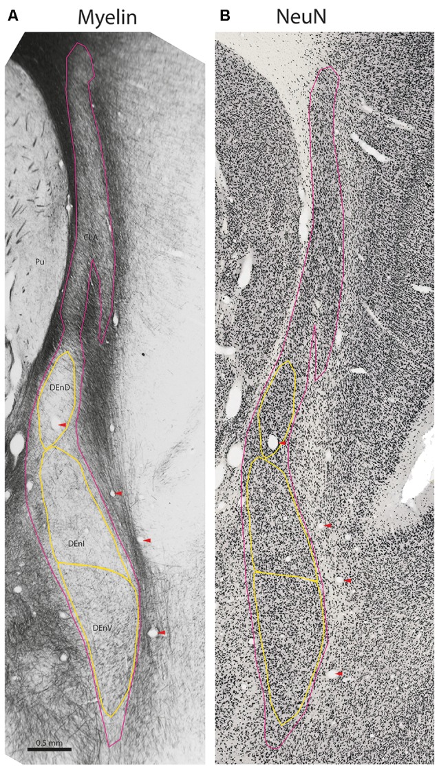
Figure 2.

Internal subdivisions of the claustrum complex in the marmoset. Adjacent coronal sections stained for myelin (Gallyas, 1979) and the neuronal marker NeuN (Atapour et al., 2018), respectively. The observed internal boundaries of the claustrum complex, including the proposed subdivisions of the DEnD, are outlined in yellow (A). Note that the sparse myelin in the DEnD region corresponds to an area of relatively high-density NeuN staining (B), but the DEnI and DEnV boundaries are not well resolved in NeuN, which was consistent across the sections examined in this study. Red arrowheads indicate blood vessels used for registration of adjacent stained sections. Case CJ167, approximate A-P +10.3 (Paxinos et al., 2012). CLA, insular claustrum; DEnD, DEnI, DEnV, dorsal, intermediate and ventral subdivisions of the dorsal endopiriform, respectively; Pu, putamen.
