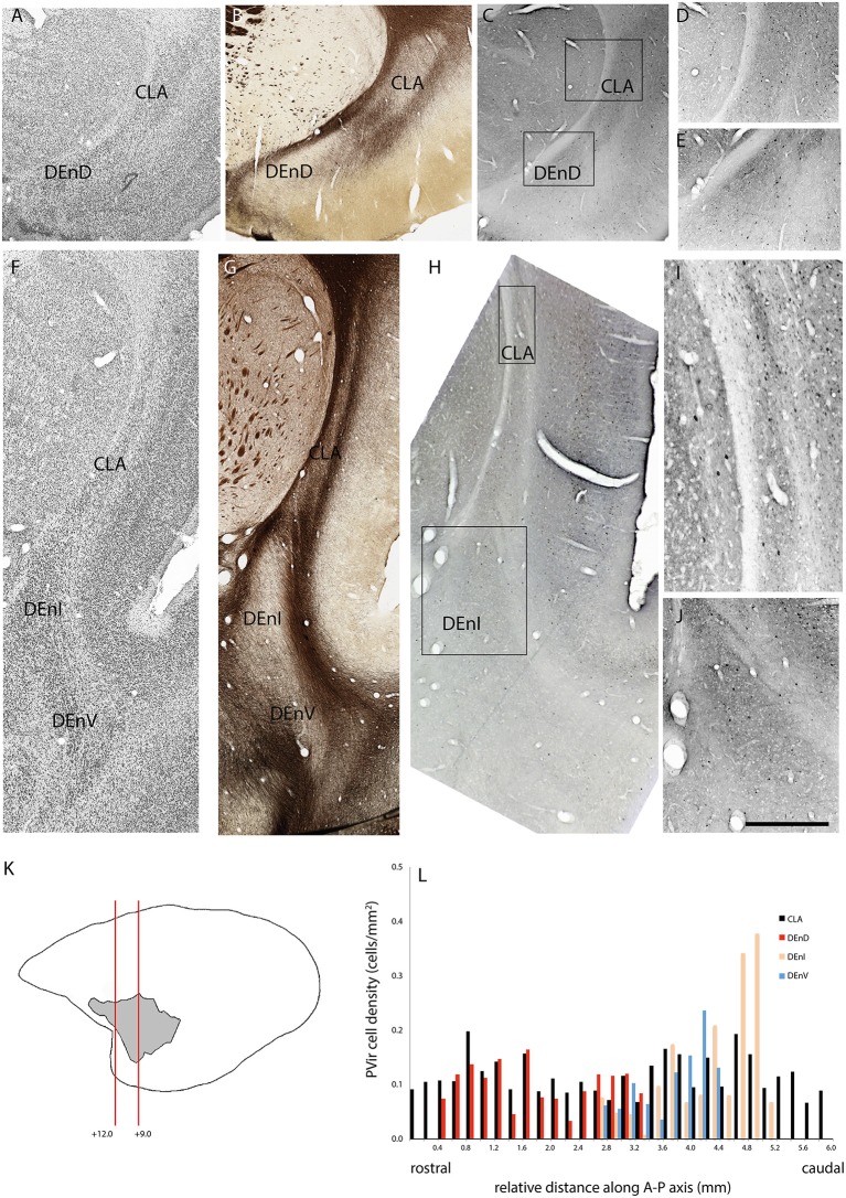
Figure 5.
Adjacent rostral- (A–E) and mid- (F–J) coronal sections of the claustrum complex showing Nissl (A,F); myelin (B,G); and parvalbumin immunoreactive (C–E,H–J) staining in the CLA and DEnD subdivisions at each level from case F1882. (K,L) Figure annotations and conventions as in Figure 4.

