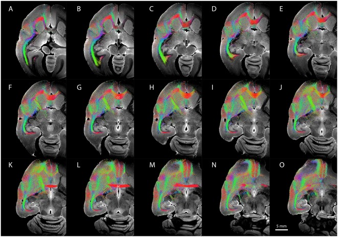
Figure 6.
Diffusion-weighted imaging (DWI) tractography showing streamlines from the entire claustrum complex in case CJ197. (A–O) Horizontal sections showing fiber orientation distribution (FOD) for left cerebral hemisphere. Note high streamline density in orbital prefrontal cortex (A–F), insular cortex (D–H), and auditory cortex (F–K) lateral to the corpus callosum (labeled cc in G), and in medial prefrontal cortex (K–O) rostral to the anterior commissure (labeled ac in L).

