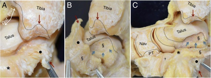
Fig. 3.
Detailed observation of the joint capsule in the medial ankle. a The medial part of the capsule appeared fibrous (black circle). There is a clear qualitative difference between the anterior fatty tissue (asterisk) and the medial fibrous tissue. Red arrows indicate the corresponding bone and detached capsule. b The fibrous capsule occupied the space of the intercolliculus groove like a mortise joint. The corresponding side of the fibrous capsule (section mark in Fig. 3b) widely attached to the depressed area of the medial side of the talus (blue arrows), which formed an ellipse. c The posteromedial fibrous capsule extended distally, and changed to cartilaginous tissue (dagger). The cartilaginous tissue covered the talus head, and attached to the proximal portion of the PT tendon insertion (white dotted line). Similar marks represent the attachments of the capsule and corresponding parts of the capsule. Prox, proximal; Dist, distal; Nav, navicular; PT, insertion of the posterior tibialis tendon

