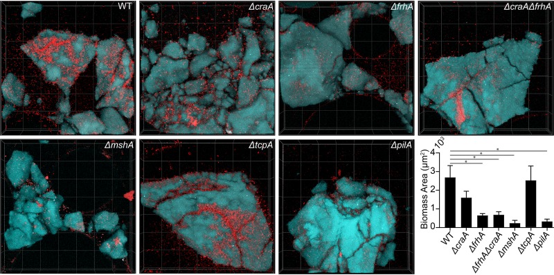
FIG 9.
ΔcraA and ΔfrhA mutants in V. cholerae O1 El Tor A1552 show attachment defects on chitin. All images are top down 3D renderings with dimensions of 212 μm by 212 μm by 33 μm (length by width by depth); red indicates cellular biomass and cyan shows the chitin particles. Quantification of the biomass in of each strain was performed after 48 h of growth of 3 biological replicates. Wilcoxon signed-ranks tests were performed to compare each mutant to the wild type (WT). *, P < 0.05. All other mutants shown were found to not be significantly different from WT.

