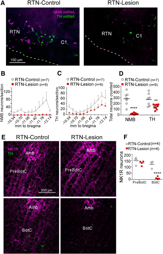
Figure 2.
Histological analysis of RTN lesions. A, NMB mRNA-expressing RTN neurons (magenta) and Th mRNA-expressing C1 neurons (green) in a transverse section (bregma level −11.6 mm; all bregma references after Paxinos and Watson, 1998) from a saline (left, RTN-Control) and SSP-Saporin (SSP-SAP, right, RTN-Lesion) treated rat. White dashed line indicates the ventral surface of the medulla oblongata. There is absence of NMB+ neurons in the RTN and preservation of C1 catecholaminergic neurons. B, Bilateral counts of NMB+ neurons in the RTN by bregma level. C, Bilateral counts of Th+ neurons by bregma level. D, Total bilateral counts of NMB and TH neurons throughout the RTN in saline and SSP-SAP treated rats (unpaired two-tailed Student's t test, for NMB: t(14) = 7.1, p < 0.0001 (****); for TH: t(14) = 3, p = 0.0093 (**). E, Neurokinin 1 receptor (NK1R, magenta) and TH (green) expression in transverse sections of the Bötzinger Complex (BötC) (top, bregma level −11.8 mm) and Pre-Bötzinger Complex (Pre-BötC) (bottom; bregma level −12.8 mm) in a saline rat (left) and SSP-SAP (right) treated rat. Amb, Nucleus ambiguus. F, Bilateral counts of NK1R-immunoreactive neuronal cell bodies in a single transverse section containing the BötC and pre-BötC. Two-way ANOVA, effect of region (pre-BötC vs BötC), F(1,6) = 114.7, p < 0.0001; effect of lesion, F(1,6) = 23.1, p = 0.003; interaction between lesion and regions, F(1,6) = 43.6, p = 0.0006. Bonferroni-corrected post hoc t test: effect of lesion in pre-BötC, t(12) = 1.2, p = 0.48; effect of lesion in BötC, t(12) = 7.3, p < 0.0001 (****).

