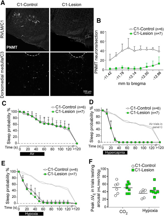
Figure 5.
C1 neuron ablation has no effect on arousal or ventilatory stimulation during hypercapnia or hypoxia. A, Transverse sections showing phenylethanolamine-N-methyl transferase-immunoreactive neurons in the rostral ventrolateral medulla (RVLM) (C1 region, top, bregma level −12.0 mm) and dorsomedial medulla (C3 region, bottom, bregma level −11.4 mm) in a TH-Cre rat injected with AAV2-DIO-hM3D(Gq)-mCherry (C1-Control, left) or AAV5-Flex-taCasp3-TEVp (C1-Lesion, right). B, Counts of PNMT-immunoreactive cells in the ventrolateral medulla by bregma level (N = 6 for C1-Control, N = 7 for C1-Lesion). C, Sleep probability curves during air-to-air trials (C1-Control: 11 ± 3 trials/rat; C1-Lesion: 11 ± 2 trials/rat). Repeated-measures two-way ANOVA; interaction between C1-Lesion and time, F(13,143) = 0.4, p = 0.98; time effect, F(13,143) = 106.9, p < 0.0001; lesion effect, F(1,11) = 0.01, p = 0.91. Light gray represents air-to-air trials. D, E, Error bars. D, Sleep probability curves during CO2 trials (C1-Control: 11 ± 2 trials/rat; C1-Lesion: 13 ± 5 trials/rat). Repeated-measures two-way ANOVA; interaction between lesion and time, F(13,143) = 0.12, p = 0.99; time effect, F(13,143) = 361.9, p < 0.0001; lesion effect, F(1,11) = 0.02, p = 0.88. E, Sleep probability curves during hypoxia trials (C1-Control: 12 ± 4 trials/rat; C1-Lesion: 13 ± 4 trials/rat). Repeated-measures two-way ANOVA; interaction between C1-Lesion and time, F(13,143) = 1.4, p = 0.17; time effect, F(13,143) = 268.1, p < 0.0001; lesion effect, F(1,11) = 1.3, p = 0.27. F, Peak ΔVE during trials testing arousal for C1-Lesion (N = 7) versus C1-Control (N = 6) rats (CO2, unpaired t(11) = 0.22, p = 0.83; hypoxia, unpaired t(11) = 1.2, p = 0.28).

