Figure 4.
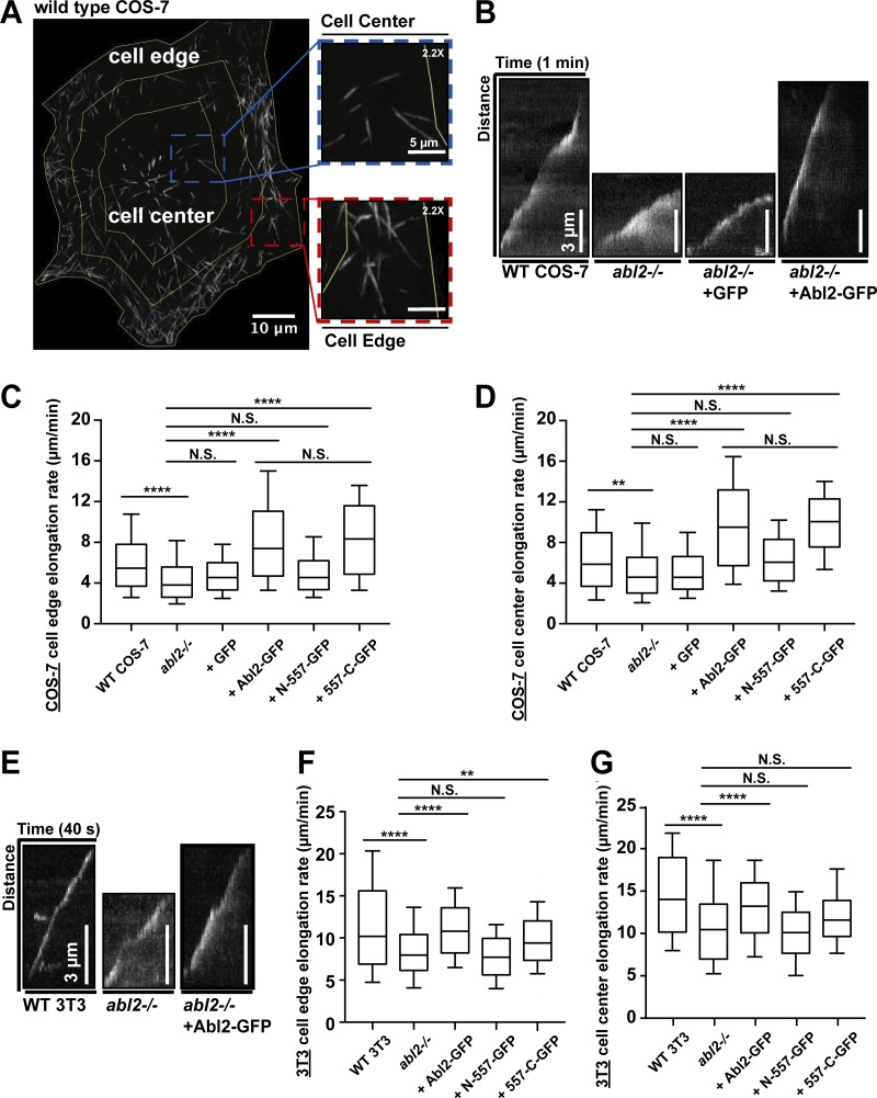
Abl2 is required for normal MT growth in cells. Maximum intensity projection of mCherry-MACF43 time-lapse showed single MT tracks in a COS-7 cell. MT growth events in the inner one half of the cell, centered at the MTOC, were categorized as being within the cell center, and those in the outermost one quarter region were categorized as being within the cell edge. (B) Kymographs of the cell edge MT plus-tip growth in WT and abl2−/− COS-7 cells, and abl2−/− COS-7 cells re-expressing GFP or Abl2-GFP; (E) in WT and abl2−/− 3T3 cells, and abl2−/− 3T3 cells re-expressing Abl2-GFP, which are also shown in Fig. S3 E as references. Quantifications of MT plus-tip growth at the cell center and the cell edge in (C and D) COS-7 or (F and G) 3T3 WT, abl2−/−, and abl2−/− re-expressing Abl2-, N-557-, 557-C-GFP cells. n ≥ 150. **, P < 0.01; ****, P < 0.0001. N.S., not significant.

