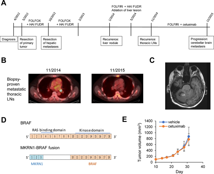
Fig. 4. Class 2 mutations can cause secondary resistance to EGFR inhibitors.
(A) Timeline of patient’s treatment history, (B) Representative images from PET/CT showing biopsy-proven, hypermetabolic metastatic thoracic lymphadenopathy at the start of FOLFIRI/cetuximab treatment and after one year of treatment, (C) Representative MRI image showing new cerebellar metastasis, (D) Schema of the MKRN1-BRAF fusion identified on sequencing the cerebellar metastasis, (E) Growth curve of mice bearing the PDX with MKRN1-BRAF fusion treated with vehicle or cetuximab. Five mice were treated in each group and standard deviations are indicated with error bars.

