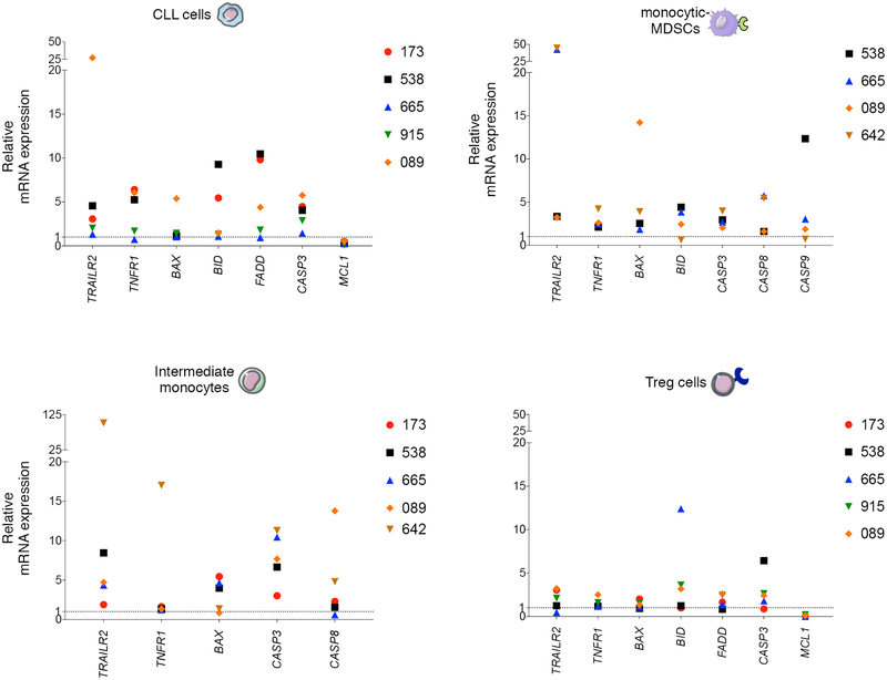
Figure 2. Trabectedin-induced cell death mechanisms in selected primary lymphoid and myeloid cells from CLL patients.
Relative mRNA expression of TRAILR2, TNFR1, BAX, BID, FADD, CASP 3, CASP 8, CASP 9, and MCL1 in hCD19+CD5+ human primary CLL cells, CD14+HLADRlow/- M-MDSCs, intermediate monocytes and CD4+CD25+CD127low/- Tregs separated by FACS from PBMCs (n=6, patients 173, 538, 665, 915, 089, 642, Supplementary Table S2) and plated in 6-well plates alone or with 0.01 μM of trabectedin for 15 h. Three technical replicates were analyzed for each sample. Data were normalized to β-actin expression. Gene expression was determined by calculating the difference (ΔCt) between the threshold cycle (Ct) of each gene and that of the reference gene and was expressed as the mean of 3 replicates ± SEM. Then the relative quantification values were calculated as the fold change expression of the gene of interest over its expression in the selected cell type reference sample, i.e., the untreated sample (considered as the calibrator sample), by the formula 2 – ΔΔCt. Finally the treated sample relative mRNA expression was normalized to the vehicle. Samples with undetermined Ct values are not included.

