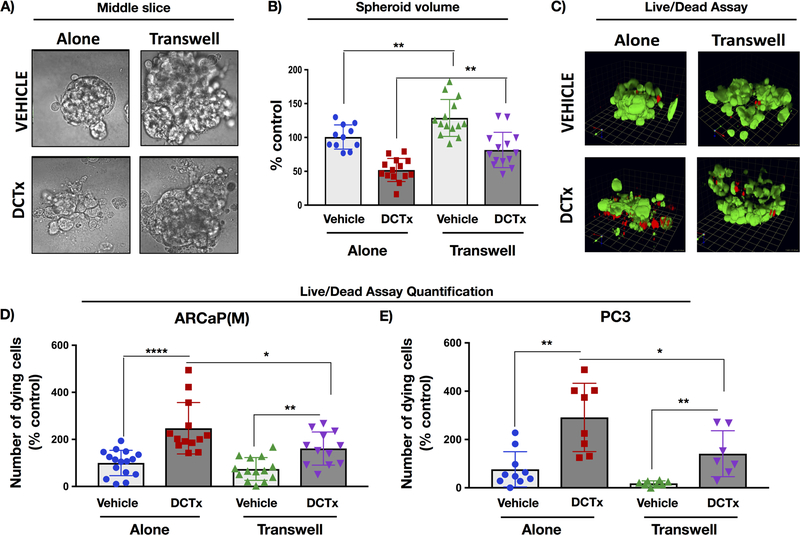
Figure 1. Interaction with adipocytes reduces sensitivity of 3D spheroids from ARCaP(M) and PC3 cells to docetaxel (DCTx).
3D cultures of ARCaP(M) grown in Transwell co-culture with bone marrow adipocytes in the absence or presence of 10nM DCTx. A: DIC images of a slice through the middle of 3D spheroid; B: Quantification of total spheroid volume; C: 3D reconstruction of Live/Dead assay results from ARCaP(M) spheroids grown alone or in Transwell with marrow adipocytes and treated with vehicle (EtOH) or 10nM DCTx; green: Calcein AM-positive live cells; red: ethidium homodimer–positive dead cells; Quantification of ethidium–positive (dead) ARCaP(M) (D) and PC3 (E) cells per total spheroid volume shown as percent control; * p<0.05; **p<0.01; ****p<0.0001.

