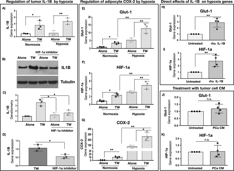
Figure 6. COX-2 levels in adipocytes and IL-1β levels in PCa cells are sensitive to hypoxia.
A: Gene expression of IL-1β in PC3 cells cultured alone or in Transwell with adipocytes under normoxic or hypoxic conditions; B: IL-1β protein levels in PC3 cells cultured alone or in Transwell and in the absence or presence of 5μM HIF-1α inhibitor CAY10585; representative blot is shown; C: IL-1β densitometry normalized to tubulin; data represent the mean+/− SD from 3 separate experiments; D: IL-1β gene expression in PC3 cells grown in Transwell cultures in the absence or presence of 5μM CAY10585. Gene expression of GLUT1 (E), HIF-1α (F) and COX-2 (G) in bone marrow adipocytes cultured alone or in Transwell with PC3 cells under normoxic (21% O2) or hypoxic (1% O2) conditions. Data are shown as the mean of 3 biological replicate experiments. Gene expression of GLUT1 (H) and HIF-1α (I) of adipocytes upon treatment with recombinant IL-1β. Gene expression of GLUT1 (J) and HIF-1α (K) of adipocytes upon treatment with media conditioned by PCa cells; +/− SD. * p < 0.05; ** p < 0.01; n.s. -not significant.

