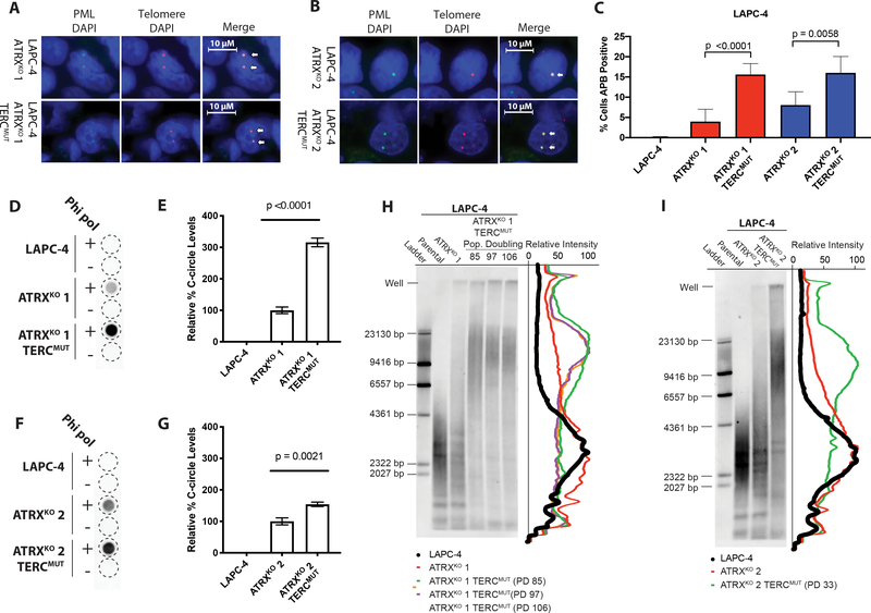
Figure 4.
APBs, C-circles, and telomere lengths increase in LAPC-4 ATRXKO clones lacking telomerase activity. Representative images of telomere FISH (red) and PML immunoblotting (green) from (A) LAPC-4 ATRXKO 1 and LAPC-4 ATRXKO 1;TERCmut; (B) LAPC-4 ATRXKO 2 and LAPC-4 ATRXKO 2;TERCmut. Arrows indicate examples of ALT-associated PML Bodies (APBs). Cells were scored for APBs in (C) LAPC-4 ATRXKO 1;TERCmut and LAPC-4 ATRXKO 2;TERCmut. P-values were determined using two-sided t-tests. C-circle assay of (D) LAPC-4 ATRXKO 1;TERCmut and (F) LAPC-4 ATRXKO 2;TERCmut. The relative quantity of C-circle was assessed from blots of (E) LAPC-4 ATRXKO 1;TERCmut and (G) LAPC-4 ATRXKO 2;TERCmut. P-values were determined using unpaired two-sided t-tests. Representative telomere restriction fragment (TRF) southern blots of (H) LAPC-4 ATRXKO 1;TERCmut and (I) LAPC-4 ATRXKO 2;TERCmut. Intensity traces of the distribution of TRFs is shown. PD = population doubling.

