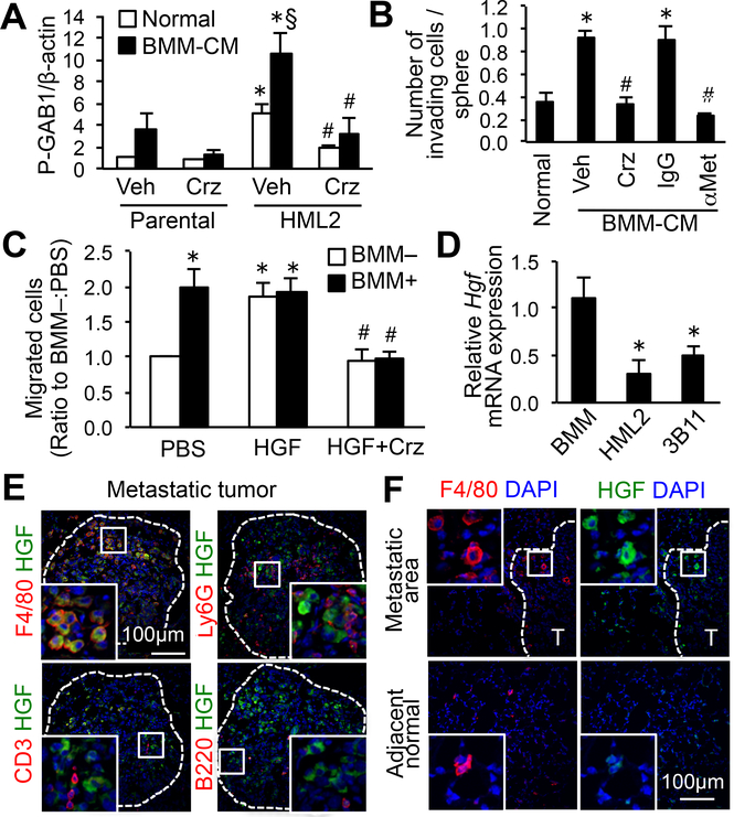
Figure 3. Macrophage-derived HGF activates MET signaling in metastatic cancer cells.
(A) Phospho-GAB1 relative to β-actin in parental and HML2 cells stimulated with normal medium or BMM-conditioned medium (BMM-CM) for 4 hours with MET inhibitor crizotinib (Crz) or vehicle (n=5). *P<0.05 versus parental; §P<0.05 versus normal; #P<0.01 versus vehicle, Student’s t test. (B) Average number of invading cells in E0771-HML2 spheroids cultured with normal medium or BMM-CM for 48 hours in the presence of Crizotinib, vehicle, blocking antibody to MET (αMet), or control IgG (n=9). *P<0.01 versus normal; #P<0.01 versus vehicle/IgG controls, Student’s t test. (C) Number of extravasated E0771-HML2 cells in vitro. Cancer cells were cultured for 36 hours in the presence or absence of BMMs with or without HGF and crizotinib (n=6). *P<0.01 versus PBS:BMM–; #P<0.01 versus HGF, Student’s t test. (D) Relative Hgf mRNA expression in BMMs, E0771-HML2, and 3B11 endothelial cells (n=6). *P<0.01 versus BMMs, Student’s t test. (E,F) Expression of HGF and markers for macrophages (F4/80), neutrophils (Ly6G), T cells (CD3), or B cells (B220) in the lungs of animals with metastatic E0771 tumors (T), and macrophages in normal adjacent tissue (20x magnification). Dotted line indicates a border of metastatic foci (n=3). Scale bar: 100 μm; Square indicates enlarged area at 60x magnification.

