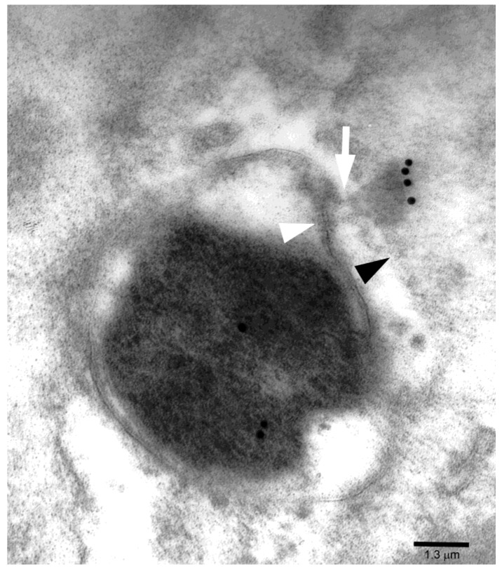
Figure 3.
Another intercellular bacterium shows CagA immunogold in its core as well as on the cytoplasmic front of a relatively dense focal structure crossing the epithelial membrane (black arrowhead) while retaining structural connection (white arrow) with bacterial outer membrane (white arrowhead).

