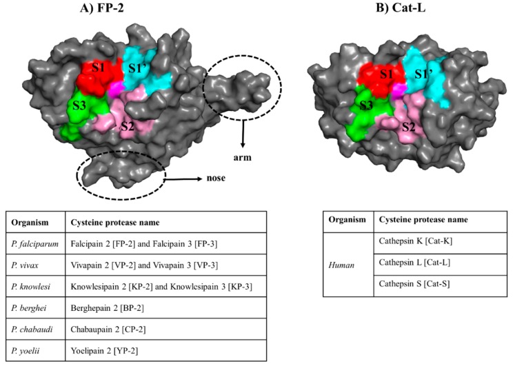
Figure 1.
The general structural fold of (A) falcipains (FPs) and plasmodial homologs and (B) human cysteine proteases. The different subsites forming the “trench-like” active pocket are shown in red (S1), pink (S2), green (S3) and cyan (S1′). The central catalytic Cys residue is colored in magenta. The unique structural features (nose and arm) found only in plasmodial proteases are marked with a broken line. Tables present the name of the homologous FP-2 and FP-3 proteins from other Plasmodium species as well as the human host.

