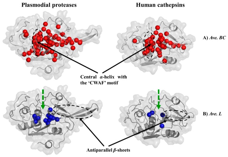
Figure 8.
Key communication hubs in plasmodial proteases and their human homologs. Structural mapping of residues with significantly high average BC (A, red) and low average L (B, blue) values in plasmodial proteases and human cathepsins in both ligand-bound and ligand-free states. Active site location is shown by the thick green broken line.

