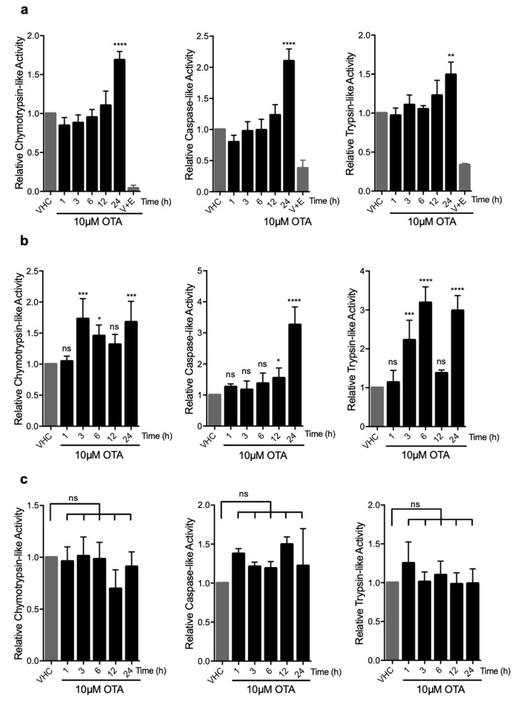
Figure 4.
OTA elevates the enzymatic activities in 26S proteasome in HK-2 and WT MEF cells but not in autophagy-deficient Atg5-/- MEF cells. (a) HK-2 cells, (b) WT MEF cells, and (c) Atg5-/- MEF cells were treated with 10 µM OTA for indicated time periods or vehicle (0.1% v/v Et-OH) for 24 h. HK-2 cells were additionally treated with 10 µM OTA and proteasome inhibitors (250 nM Epoxomicin (E) and 5 µM VR23 (V) for 24 h. Lysates containing 26S proteasome were extracted from the cells and proteasome activities were measured by using Proteasome-Glo® assay (Promega, USA). Suc-LLVY-Glo, Z-nLPnLD-Glo, and Z-LRR-Glo substrates were used to measure chymotrypsin-, trypsin-, and caspase-like activities, respectively. The results were normalized to PSMB5, PSMB6, and PSMB7 protein levels for chymotrypsin-, caspase-, and trypsin-like activities, respectively and expressed as relative to vehicle (VHC) control (ns: Nonsignificant, * p < 0.05, ** p < 0.01, *** p < 0.001, **** p < 0.0001).

