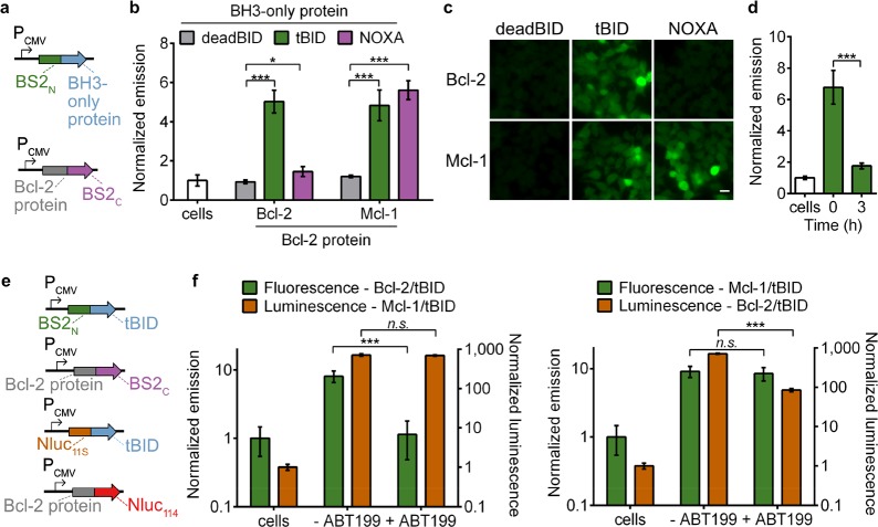
Figure 3.
Split BS2 can detect Bcl-2 family PPIs and inhibitor engagement. (a) Vector system to test Bcl-2 split esterase PPI detection. (b) HEK293T cells cotransfected with the plasmids shown in part a were incubated with fluorescein-CM2 and analyzed for fluorescence by a plate reader. The normalized emission for interactions between deadBID/Bcl-2 protein (gray), tBID/Bcl-2 protein (green), and NOXA/Bcl-2 protein is shown. HEK293T control cells (white) were similarly analyzed. (c) HEK293T cells cotransfected and incubated with fluorescein-CM2 as in part b were analyzed by fluorescence microscopy. (d) HEK293T cells cotransfected with BS2N-fused tBID and Bcl-2-fused BS2C were treated with DMSO (time = 0) or ABT199 for 0–3 h (green) followed by incubation with fluorescein-CM2 and analyzed for fluorescence by a plate reader. HEK293T control cells (white) were similarly analyzed. (e) Vector system to simultaneously detect two PPIs and Bcl-2 inhibition. (f) HEK293T cells were cotransfected with the split esterase plasmids or split Nluc plasmids shown in part e. The two cell populations were mixed and treated with ABT-199 or a DMSO control. After 24 h, the cells were incubated with fluorescein-CM2 and analyzed (green). Immediately after analysis, furimazine was added, and the cells were reimaged (orange). The Bcl-2/tBID interaction was selectively blocked and detected with the esterase reporter (left) or Nluc reporter (right). HEK293T control cells were similarly analyzed. Error bars are the standard deviation for n = 4 (b), n = 6 replicates (d), and n = 8 replicates (f). Unpaired t test; *P < 0.01, ***P < 0.0001. Scale bars shown are 20 μm.

