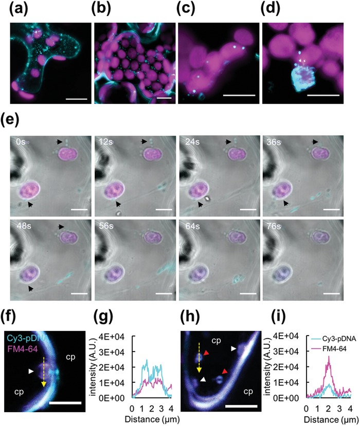
Figure 5.

Targeting pDNA delivery to the plastid in a plant cell using a combination of a CTP and CPP. a,b) Internalization of the clustered pDNA/CTP/CPP complexes into plant cells. CLSM images of an N. benthamiana leaf at 2 h after transformation with Cy3‐labeled pDNA/CTP/CPP complexes. CLSM observation of a) the spongy mesophyll cells and b) palisade mesophyll cells reveals the presence of fluorescent Cy3‐labeled pDNA molecules (cyan) delivered using a CTP/CPP complex into cells. Autofluorescent signals of chlorophyll are shown in magenta. c,d) Targeting of pDNA molecules to the plastids. c) At 2 h post‐infiltration, the Cy3‐labeled pDNA/CTP/CPP complexes (cyan) were found to colocalize with the plastids (magenta) in the plant cells. d) One of several plastids extensively covered by the Cy3‐labeled pDNA/CTP/CPP complexes. e) Time‐course observation of Cy3‐labeled pDNA/CTP/CPP complexes delivered to the chloroplasts. Arrowheads indicate the original positions of the complexes before approaching the chloroplasts. f–i) Vesicle formation in the Cy3‐pDNA/CTP/CPP complex‐transfected cells. Colocalization of the Cy3‐pDNA/CTP/CPP complexes (cyan) with the plasma membrane‐staining fluorescent dye FM4‐64 (magenta) observed by CLSM imaging at 2 h post‐infiltration. The white arrowhead indicates the membrane‐bound vesicles in (f) and (h), while the red arrowheads indicate the free vesicles in the cytoplasm (cp) in (h). g,i) Fluorescent profiles (represented by the yellow dashed line across the vesicles) indicate the colocalization of the clustered pDNA/CTP/CPP complexes with membrane‐bound vesicles in (f) or free vesicles in (h). The plant cell membrane was stained with FM4‐64 (magenta). Scale bars = 10 µm in (a) and (b), 5 µm in (c)–(e), and 4 µm in (f) and (h).
