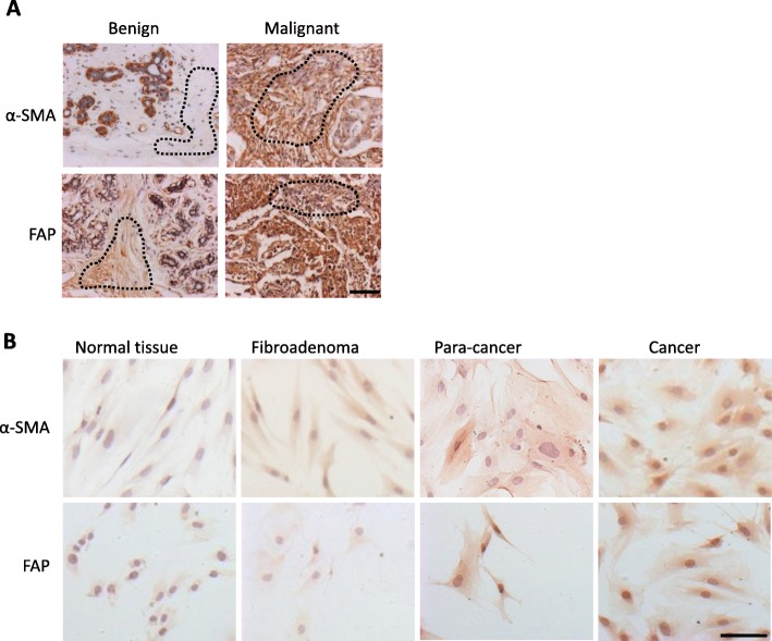
Fig. 1.
α-SMA and FAP expression in benign or malignant human breast tissues and isolated fibroblasts. a Immunohistochemical staining of human breast tissue arrays. Dotted lines indicate the stromal regions. Typical positive samples of malignant stromal tissues were selected and showed higher intensity staining (brown) with α-SMA and FAP antibodies than that of benign tissues. Scale bar, 100 μm. b Immunocytochemical staining of α-SMA and FAP in primary fibroblasts isolated from different patients with benign or malignant breast diseases. Scale bar, 100 μm

