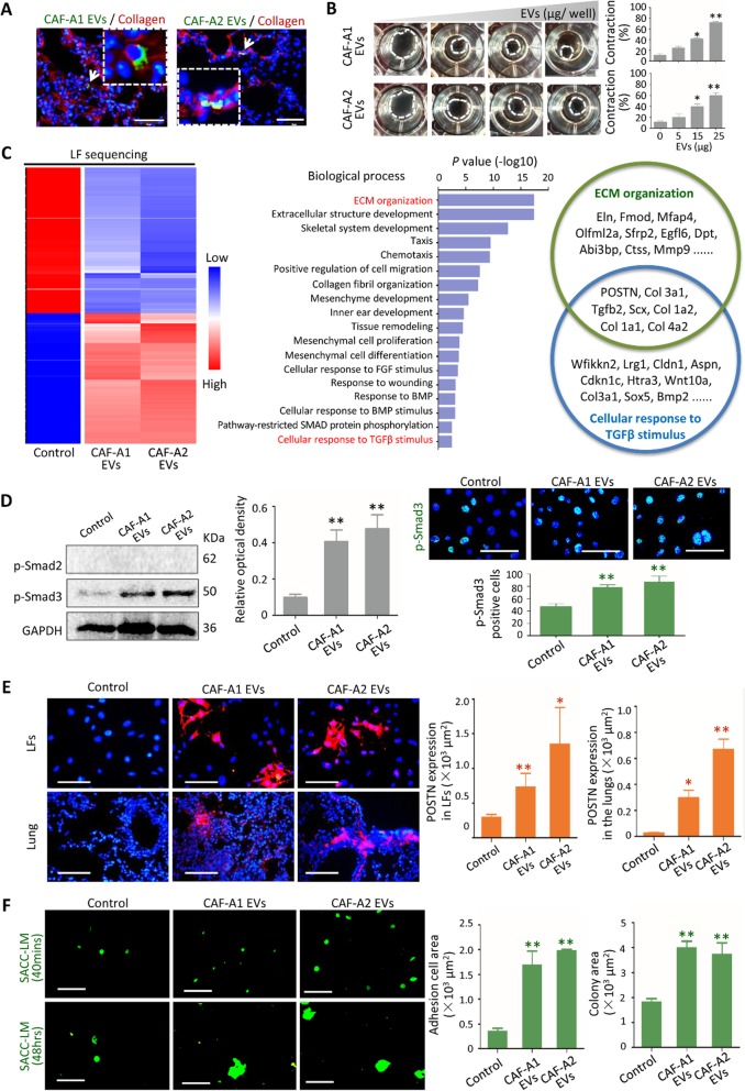
Fig. 2.
CAF EVs activated LFs via TGF-β signaling pathway. a CAF-A1/A2 EVs were labeled with PKH67 (green) and the lung tissue sections were stained with Collagen I antibody (red). Scale bar = 50 μm. b Collagen gel contract assay using EVs from CAF-A1 and CAF-A2. EV concentrations tested were 0, 5, 15, 25 μg/well. c Heatmap of RNA sequencing. Enriched Biological Processes with GO term using differentially expressed genes were listed. POSTN was the top one among the enriched gene in both ECM Organization and Cellular Response to TGF-β Stimulus Biological Processes. d Expression of p-Smad2 and p-Smad3 in LFs with or without CAF EV pre-treatment assessed by western blot and immunofluorescent staining. Scale bar = 100 μm. e POSTN expression in LFs in vitro and in the lung tissues of mice (n = 5 per group) treated with CAF-A1/A2 EVs for 3 days. Scale bar = 100 μm. f Images and quantization of adhesion and proliferation of SACC-LM-GFP cells (green) on LFs treated with EVs from SACC-LM, CAF-A1 and CAF-A2. Scale bar = 100 μm. * P < 0.05, ** P < 0.01, *** P < 0.001

