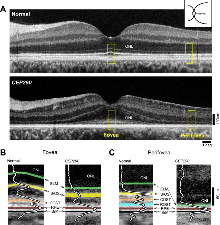
Figure 3.
Retained photoreceptor nuclei with abnormal segments in CEP290-LCA. (A) OCT scans along the horizontal meridian through the fovea in a normal subject, and a CEP290-LCA patient. Images were obtained with a clinical ultrahigh resolution SDOCT system (Bi-μ; Kowa Company, Ltd.). Hyposcattering layer corresponding to the ONL is shown. Inset upper right shows location of scan. Yellow boxes outline foveal and perifoveal regions shown in (B, C). (B, C) Magnified views of the outer retina at foveal and temporal perifoveal locations demonstrating differences in the layers distal to the ONL. Overlaid are the longitudinal reflectivity profiles (LRPs). Hyperscattering signals highlighted as follows: green, ELM; yellow, IS/OS, near the junction of inner and outer segments; orange, COST, near the interface of cone outer segment tips and RPE contact cylinder (also called the interdigitation zone); cyan, ROST, near the interface between rod outer segment tips and RPE apical processes; brown, RPE, near the RPE cell bodies; and black, BrM, Bruch membrane. Figure courtesy of Alexander Sumaroka (Scheie Eye Institute, University of Pennsylvania).

