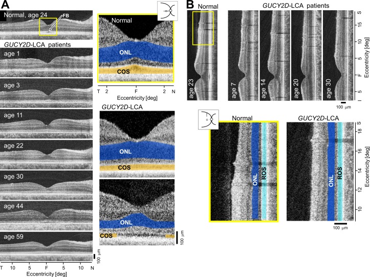
Figure 9.
Retinal structure at the fovea and perifovea in GUCY2D-LCA. (A) Cross-sectional OCT scans along the horizontal meridian through the fovea (F) in a normal subject and seven GUCY2D-LCA patients, ranging in age from 1 to 59 years. Enlarged central scans (yellow box) in a normal subject and two GUCY2D-LCA patients are also shown. The ONL is highlighted in blue and the COS layer is in orange. The upper image from a patient illustrates a thinned COS layer but normal ONL. The lower image is from a patient with reduced ONL thickness and an interrupted COS layer, suggesting a central absence of COS. FB, foveal bulge. (B) OCT scans along the vertical meridian including the fovea and continuing into the superior perifoveal region in a normal subject and four patients with GUCY2D-LCA ranging in age from 7 to 30 years. Enlarged scans (yellow box) in a normal subject and a patient showing comparable ONL (blue) and rod outer segment layer (light blue) thickness. Figure courtesy of Alexander Sumaroka (Scheie Eye Institute, University of Pennsylvania).

