Figure 3:
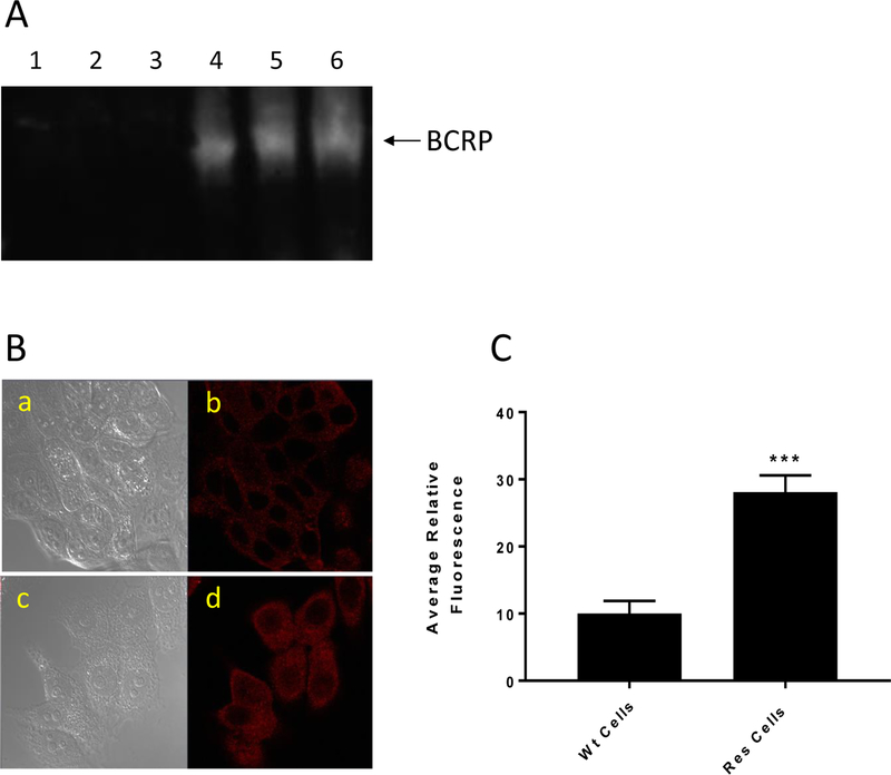
The Western blot analysis for BCRP in MCF-7 and MCF-7/MX cells (A). Lanes 1, 2, 3 and 4, 5, 6 represent 5,10 and 20 μg proteins from MCF-7 and MCF-7/MX tumor cells, respectively. (B) Confocal microscopy studies for BCRP in MCF-7 and MCF-7/MX tumor cells. a, WT MCF-7 cells without BCRP antibody; b, in the presence of the antibody; c, MCF-7/MX cells without the antibody; and d, MCF-7/MX cells in the presence of the antibody. (C) quantifications of cellular fluorescence of BCRP.*** p values ≤ 0.001.

