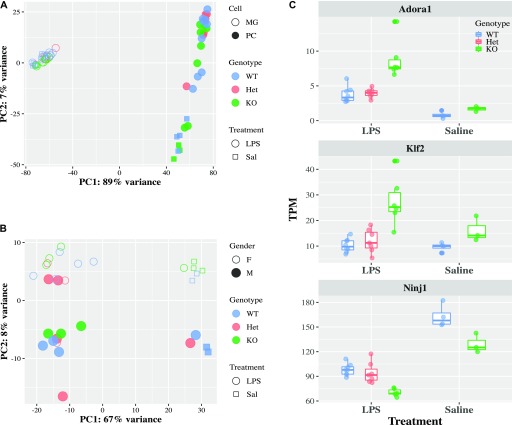
Figure 3. Loss of Cx3cr1 does not affect microglial transcriptional response to LPS.
2-mo-old Cx3cr1+/+ (WT, n = 7), Cx3cr1+/eGFP (Het, n = 6), or Cx3cr1eGFP/eGFP (KO, n = 6) mice were injected with 1 mg/kg LPS i.p. (or saline control, four WT and three KO animals only). Twenty-four hours later, peritoneal cells were collected, and microglia were sorted from isolated brains. Both cell populations were used for RNA-seq. (A) PCA of all samples. Each dot within a cell type represents an individual animal, but there are matched microglia and peritoneal cells from the same animal. Samples, coded based on the cell type (open or solid fill), genotype (color), and treatment (symbol), separate by cell type and treatment. (B) The PCA of microglia only shows separation by treatment, but not genotype in LPS-injected mice. Samples are coded by gender (open or solid fill), genotype (color), and treatment (symbol). (C) Expression of selected genes that are modulated by Cx3cr1 genotype after LPS injection.

