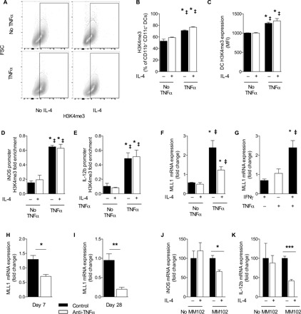
Fig. 4. TNFα-programmed DC1 phenotype and gene promoters associated with increased H3K4me3 and MLL1 are necessary for TNFα programming of DC1.

DC1-polarized BMDCs were treated initially with IFNγ in the presence (TNFα) or absence (No TNFα) of supplemental TNFα. (A to C) Global H3K4me3 signature was assessed by intranuclear flow cytometry between DC1, DC2, and TNFα-programmed DC1. (D and E) ChIP was performed with H3K4me3 antibodies on DC1, DC2, and TNFα-programmed DC1 with subsequent PCR using iNOS and IL-12b promoter region–specific primers. Data are presented as already normalized to β-actin. n = 3 replicates pooled from three separate experiments of 6 million DCs. For (B) to (E), *P < 0.05 between the indicated sample and DC1 and ‡P < 0.05 between the indicative sample and DC2. (F) DC1-polarized BMDCs were treated initially with IFNγ in the presence (TNFα) or absence (No TNFα) of supplemental TNFα and then challenged with IL-4, and qPCR for MLL1 was performed. *P < 0.05 relative to DC1 and ‡P < 0.05 relative to DC2. n = 18 from three separate matched experiments. (G) MLL1 was quantified by mRNA expression in DC1, in unpolarized BMDC treated only with TNFα, and in BMDC treated with TNFα and IFNγ combined. *P < 0.05 relative to DC1 and ‡P < 0.05 relative to unpolarized DC treated with TNFα alone. n = 18 from three separate matched experiments. (H and I) MLL1 mRNA was quantified from magnetically sorted CD11c+ pulmonary cells at early (H) and late (I) points of infection. *P < 0.05, **P < 0.01, and ***P < 0.001 between indicated samples; no pairing was performed. (J and K) TNFα-programmed DC1s were generated in the presence or absence of MLL1 inhibitor MM102 and then challenged with IL-4. *P < 0.05 and ***P < 0.001 between the indicated samples.
