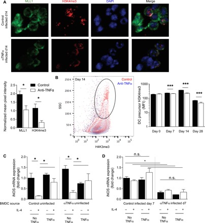
Fig. 6. TNFα is required for prepolarization of DC1 from BM throughout C. neo infection.

BM was harvested from control or αTNFα mice at the indicated times after infection. (A) Magnetically separated BM DC precursors were stained for MLL1 (green), H3K4me3 (red), and 4′,6-diamidino-2-phenylindole (DAPI) (blue). Representative images were taken at 40×. Fluorescence intensity was measured by imageJ and normalized to DAPI between images. n = 6 independent wells from separate matched experiments; three random fields were analyzed from each independent well. (B) FACS (fluorescence-activated cell sorting)–sorted DC precursors were stained for intranuclear H3K4me3; representative histograms of 14 dpi with cumulative bar graph of MFI throughout infection are shown. n = 8 from separate matched experiments. ***P < 0.001. (C and D) BMDCs were matured for 7 days ex vivo with GM-CSF from BM of uninfected control and αTNFα mice (C) or 7 dpi control and αTNFα mice (D) and then treated as in Fig. 1. DC1 gene stability was assessed. Data are normalized to control DC1 values within each graph. n = 6 from separate matched experiments. *P < 0.05 by ANOVA within uninfected control and uninfected αTNFα and ‡P < 0.05 by ANOVA between infected control and αTNFα. n.s., not significant.
